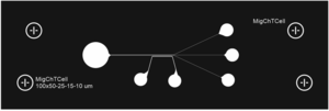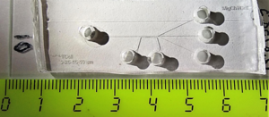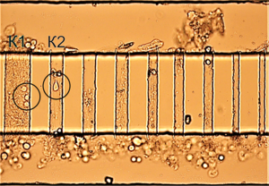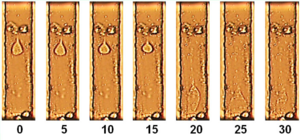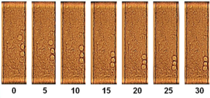

The paper presents the experimental and theoretical aspects of studying the migration of cancer cells through narrow microchannels when interacting with a chemical substance — an attractant. Chemical agents were injected directly from the respective separate reservoir via hydrostatic head. The presence of migration channels of different sizes allowed us to analyze the migration abilities inherent in the cells. Experimental studies on the migration of cancer cell lines were carried out using a microfluidic binary migration cell. It consists of two chambers 50 μm deep, connected by a series of narrow microchannels 10 μm high. The chambers are connected to the tanks from which cancer cells and activating media come. The model features smooth transitions from all tanks to microchannels to prevent accumulation of microbubbles and cells, the presence of a restrictive barrier between a chemoattractant container and a nutrient medium to reduce the effect of capillary forces, sealing the supply channels and cells with cells using a glass plate glued on top. In a series of experiments, the ability of epithelial-like cells of the metastatic prostate cancer DU145 to migrate in the migration microfluidic system developed by us was revealed. The nature of migration depends on the width of the channel and the location of the cells at the time of adding the chemical agent. In wider channels, cells adhere more slowly and are able to perform rolling movements. In narrower channels, the cells are spread over the glass. The mathematical model of concentration-capillary movement, which is a system of equations of the dynamics of an incompressible viscous fluid, written separately for the cell contents and for its environment, is considered as the basis for a theoretical study of cell migration.
cell migration,
chemotaxis,
microfluidic systems,
biological systems modeling,
prostate cancer
Experimental and theoretical aspects of studying the migration of cancer cells through narrow microchannels when interacting with a chemical substance, an attractant, are presented. Chemical agents were injected from the reservoir by a hydrostatic pump. In the laminar flow due to diffusion, a chemoattractant concentration gradient was formed, leading to cell migration.
Objective: Using migration channels of different sizes to analyze the inherent in the cells of migration features in order to build a multiphase model of active migration of cancer cells.
Method:
We used PC-3 cell lines (obtained from bone metastasis of a patient with stage IV adenocarcinoma of the prostate gland) and DU145 (obtained from metastasis of adenocarcinoma of the prostate gland to the brain). Microfluidic devices (migration cells) made by soft photolithography were used to study biological cellular systems, analyzing the movement of cancer cells in channels of comparable size to their own dimensions. Diffusion of the chemoattractant into the nutrient medium in a laminar flow leads to the formation of a concentration gradient across the channel, which should lead to the activation of cancer cells. Migration features of cancer cells were recorded using a Zeiss Axio Observer.D1 microscope in phase contrast mode. Mathematical modeling is implemented on the basis of the equations of the mechanics of multiphase media, taking into account changes in capillary forces under the influence of a chemical agent.
Results and conclusions:
A microfluidic binary migration cell has been developed, which consists of two chambers 50 μm deep each connected by a series of narrow microchannels 10 μm high. For the manufacture of a two-tier migration chamber, photomask masks were developed in vector drawings for printing on a high-resolution printer: 1) a photomask of migration channels with a thickness of 10 μm; 2) the mask of the photomask with the entrance flow and lower non-flowing channel with a thickness of 50 microns. A photo mask was made on the basis of the first mask, then a photoresist was applied on it with a layer of 50 μm. Next, on the basis of the second mask, the remaining part of the photomask of the cell 50 μm thick was fabricated. On the basis of a two-level photomask, a replica was made in a plate made of polydimethylsiloxane (PDMS), then holes were made with a diameter of 3 mm in a 5 mm thick PDMS and it was glued to a glass slide, the microcell was ready. The chambers are connected to the tanks from which cancer cells and activating media come. The features of the model are smoothed transitions from all tanks into microchannels, preventing the accumulation of microbubbles and cells, the presence of a delimiting barrier between the chemoattractant and the nutrient medium to reduce the effect of capillary forces, sealing the supply cells with a glass plate. As a basis for describing cell migration, a mathematical model of concentration-capillary movement is considered, which is a system of equations of the dynamics of an incompressible viscous fluid, written separately for the contents of the cell and for its environment.
In a series of experiments, the ability of epithelial-like cells of the metastatic prostate cancer DU145 to migrate in the microfluidic system developed by us was revealed. The nature of migration depends on the width of the channel and the location of the cells at the time of adding the chemoattractant. In wider channels, cells adhere more slowly and are able to perform rolling movements. In narrower channels, the cells are spread over the glass.
The uniqueness of the results obtained is due to the use of the developed new powerful method, which has the ability to study not only the migration features of cells, but also their deformation, which affects movement in spatially cramped conditions when exposed to different types of chemical reagents.
The resulting system of equations is designed to determine the conditions for the emergence of the velocity u of the cell drift for a given gradient of attractant in the surrounding fluid and, thus, proposed a new mechanism for the generation of migration forces, in which the chemoattractant plays the role of a surfactant.


